
Almost every patient who comes to me worried about old filler leaves with an ultrasound scan. It is quick, it happens at the same appointment, and in most cases it answers the question: where the filler is, how much is left, which layer it sits in, and whether it is touching anything it shouldn’t.
But ultrasound has limits, and part of using it well is knowing where they are. Some questions it cannot answer — because the problem is too deep, hidden behind something dense, spread across the whole face, or not actually filler at all. That is when I ask for an MRI. Not because ultrasound failed, but because the two scans see the face in different ways.
This article explains what each scan can and cannot show, how I decide between them, and what happens if you need both.
Why ultrasound comes first
High-frequency ultrasound has become the first-line imaging tool for cosmetic fillers, because it can detect and identify the common filler types directly in the skin and soft tissue.1 Each material has a recognisable appearance — hyaluronic acid looks quite different from calcium-based or permanent fillers.1,2
It also has practical strengths that matter when you are the patient:2
- It happens in real time. I can scan while you smile, frown or talk, and see how filler behaves with movement.
- Doppler maps the blood vessels. Before any needle goes in, I can see where the arteries run in that area.
- It guides treatment. If hyaluronic acid needs dissolving, ultrasound lets me place the enzyme exactly where the product is, rather than flooding the area.
- No radiation, no contrast injection, no magnet. It can be repeated as often as needed to follow progress.
- It resolves superficial layers in fine detail — the skin and the layers just beneath it, which are exactly where many filler problems sit and where MRI and CT are less precise.
I use a high-frequency Clarius scanner with Doppler. For most migrated filler, lumps, swelling questions and “how much filler do I still have?”, this is all the imaging you need.
Where ultrasound reaches its limits
1. It cannot see behind dense material
Sound waves bounce off some materials so strongly that the area behind them becomes hard to see. Calcium-based fillers cast a dark acoustic shadow; liquid silicone produces a bright, scattered “snowstorm” effect; some permanent microsphere fillers leave small comet-tail streaks.2,8 Each pattern helps identify the material, but it can also hide what lies deeper.
2. Deep tissue and the face as a whole
Ultrasound is at its best close to the surface. MRI gives excellent soft-tissue contrast deeper down, and because it images the whole face in cross-section with fixed anatomical landmarks, it shows exactly where material sits in relation to muscle, fat compartments and bone.2,4 When filler has travelled, that wider map matters: dedicated facial MRI has found filler in locations nobody expected,5 and imaging studies have shown product in deeper fat compartments than it was originally injected into.9
3. Complicated inflammation
A granuloma, an abscess, an allergic-type reaction and simple overfilling can feel almost identical under the fingers. Ultrasound separates many of these, but when the picture is mixed, MRI — often with a contrast injection — helps characterise inflammatory and granulomatous reactions and distinguish them from fluid collections and scarring.2,4,5 In one series of dedicated facial MRI scans after filler, the findings included abscesses, granulomas and allergic reactions; the same paper notes earlier work showing that clinical examination alone agreed poorly with MRI once inflammation or migration was present.5
4. When it might not be filler
This is the most important reason of all. Fillers are common incidental findings on scans, and they can both mimic disease and hide it.3,6 Radiologists use specific MRI sequences, including diffusion-weighted imaging, to help detect a problem that a filler deposit might otherwise mask.3 In one published case, a hardening cheek was assumed to be a filler complication; MRI showed the lesion was not where filler is normally placed, which prompted a biopsy — and the diagnosis turned out to be sarcoidosis, not filler at all.3
If a lump is growing, if its position doesn’t match what was injected, or if the story simply doesn’t add up, I would rather image properly than assume.
5. It depends on the person holding the probe
Ultrasound quality depends on training and experience, and findings can vary between operators.2 MRI produces a fixed set of images that a radiologist can review, compare over time and share with a surgeon if needed.
Not sure which scan you need? Start with an ultrasound assessment. If it leaves a question unanswered, I will tell you — and explain why an MRI would add something. WhatsApp the clinic: +971 52 328 6185 |
What MRI can — and cannot — tell us
MRI is not simply “a better ultrasound”. It answers different questions, and it has its own blind spots.
| Ultrasound | MRI |
Where is the filler, layer by layer? | Excellent for superficial and mid layers | Excellent, including deep planes and the whole face at once |
Which type of filler is it? | Often identifiable by its pattern | Usually not — most fillers look similar; silicone is the main exception |
Blood vessels | Doppler maps arteries in real time | Not used for this purpose |
Inflammation, granuloma, abscess | Good first assessment | Better for mixed or extensive cases, especially with contrast |
Could this be something other than filler? | May raise the question | Helps answer it, alongside biopsy where needed |
Guiding treatment | Yes — targeted, at the same visit | No — it is not a real-time tool |
Practicalities | Same appointment; no radiation or contrast | Separate booking; longer scan; standard MRI safety checks |
Two points are worth knowing. First, MRI usually cannot tell one filler brand or material from another, because most fillers have a similar water content; silicone has a distinctive signature, and calcium-based fillers are better identified by their calcifications.2,3 Second, MRI cannot guide an injection, so even when MRI is needed, treatment itself is usually planned and delivered with ultrasound.2
PET-CT is not used to assess fillers: most filler materials take up the tracer and can light up like active disease, which is a recognised source of false alarms.3,6 If you are ever having a PET-CT or MRI for another reason, tell the radiology team you have had filler.
Filler lasts far longer than most people are told
MRI has also changed what we know about how long hyaluronic acid filler stays. In a review of 33 patients scanned with MRI, filler was still visible in every one of them at least two years after their last mid-face treatment, and in one patient fifteen years later.7
That is why I scan before adding more. If the last syringe hasn’t gone, another one may be building on it — and accumulated volume is exactly the setting in which heaviness, migration and lumps tend to appear.
How I decide whether you need an MRI
Every assessment starts with your history and an ultrasound scan. I suggest MRI when one or more of these is true:
- the problem sits deeper than ultrasound can resolve clearly, or behind dense material such as calcium-based filler or silicone;
- you have had several fillers over many years, possibly including permanent ones, and we need a map of the whole face;
- inflammation is extensive, keeps returning, or doesn’t behave the way a filler reaction usually does;
- a lump is growing or sits somewhere filler would not normally be placed;
- a surgeon will be involved — for example in removing permanent filler — and needs cross-sectional images to plan.
If none of these apply, you don’t need an MRI. I would rather not add cost, time or a contrast injection when an ultrasound scan has already given us the answer.
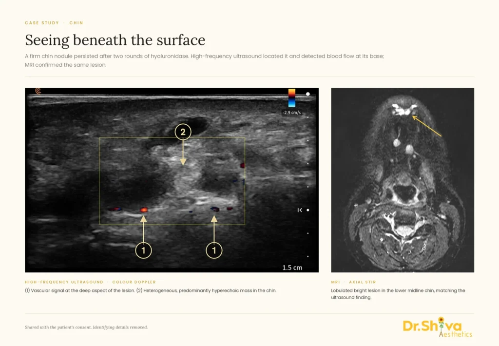
Already had an MRI elsewhere, but the report didn’t help?
Some patients come to me having already had an MRI of the face or neck, with a report that was vague, inconclusive or didn’t seem to answer their question. If that is you, please bring the original MRI images — the full study, not only screenshots — as well as the written report. Imaging centres can usually provide the complete study on a disc, a USB drive or through a download link.
An unclear report does not automatically mean the MRI itself was unhelpful. Imaging after aesthetic injections is a relatively specialised area: fillers are common incidental findings on scans, their appearance varies with the material and the time since injection, and some can resemble, or sit alongside, other conditions.3,6 The radiologist who reported your scan may not have been given your full injection history — what was injected, where and when — or the examination and ultrasound findings that make these images much easier to interpret. Without that context, a careful, cautious report is often the most accurate one possible.
With that context added, the same images can be looked at again with specific questions in mind:
- your previous facial injections and filler history, and the exact injection sites where they are known;
- what I find on clinical examination and on ultrasound;
- suspected foreign or permanent injectable material, and residual filler;
- fibrosis or scarring;
- granulomatous or inflammatory reactions;
- fluid collections or abscesses;
- relevant facial or cervical lymph nodes;
- anything that may not be filler at all and needs a different line of investigation.
Sometimes this means a new scan isn’t needed — the answer is already in the images you have. Often it isn’t only about having the MRI done; it is about who reads it, and with what information.
MRI review with our consultant radiologist
I work with Dr Abdul Rahman Alvi, MRCS, FRCR, CCT (UK), Consultant Vascular & Interventional Radiologist, who reviews imaging for our aesthetic complication patients. When you send us an MRI done elsewhere, we go through the images together: I bring your injection history, examination and ultrasound findings, and Dr Alvi reads the study with those questions in mind.
You then receive a new, official radiology report focused on your aesthetic concern — what the images show about previous filler or injected material, any inflammation, collections or scarring, and anything that needs a different line of investigation. If a finding needs an image-guided procedure, Dr Alvi can advise on that too.
|
Already have an MRI? Ask us to review it. Aesthetic-focused MRI review and official report by our consultant radiologist: AED 1,500. Send us your report first and we will tell you what we need — usually the full study on disc, USB or a download link. WhatsApp the clinic: +971 52 328 6185 |
Frequently asked questions
Is ultrasound or MRI better for filler problems? |
Neither is better in every case. Ultrasound is the first-line scan: it is immediate, maps blood vessels and guides treatment.1,2 MRI adds value for deep, extensive or unclear problems, and when something other than filler needs to be ruled out.2,3 |
Can an MRI tell me which filler I had? |
Usually not. Most fillers look similar on MRI; silicone is the main exception.2,3 Ultrasound is often more helpful for identifying the type of material.1,2 |
Will I need a contrast injection? |
Not always. Contrast is mainly used when inflammation, a granuloma or an abscess needs to be characterised.4 The radiology team will discuss it with you if it is needed. |
Can I have an MRI if I have had filler? |
Yes. Fillers are a common incidental finding on routine scans.3 The usual MRI safety questions still apply — for example pacemakers and some metal implants.2 Always tell the team you have had filler so it isn’t mistaken for something else. |
Why would I need an MRI if you can already see the filler on ultrasound? |
Seeing filler is not the same as seeing everything around it. MRI shows deep planes, the whole face at once, and whether a change might not be filler at all.2,3,5 |
How long does hyaluronic acid filler really last? |
Longer than the six to twelve months often quoted. An MRI study found hyaluronic acid in every patient at least two years after their last mid-face treatment, and up to fifteen years in one case.7 |
This article is for education and does not replace a consultation. Imaging decisions are made individually after assessment.
References
- Wortsman X. Identification and complications of cosmetic fillers: sonography first. J Ultrasound Med. 2015;34(7):1163–1172. doi:10.7863/ultra.34.7.1163
- Beiu C, Popa LG, Bălăceanu-Gurău B, Iliescu CA, Racoviță A, Popescu MN, Mihai MM. Personalization of minimally-invasive aesthetic procedures with the use of ultrasound compared to alternative imaging modalities. Diagnostics. 2023;13(23):3512. doi:10.3390/diagnostics13233512
- Mundada P, Kohler R, Boudabbous S, Toutous Trellu L, Platon A, Becker M. Injectable facial fillers: imaging features, complications, and diagnostic pitfalls at MRI and PET CT. Insights Imaging. 2017;8(6):557–572. doi:10.1007/s13244-017-0575-0
- Di Girolamo M, Mattei M, Signore A, Grippaudo FR. MRI in the evaluation of facial dermal fillers in normal and complicated cases. Eur Radiol. 2015;25(5):1431–1442. doi:10.1007/s00330-014-3513-2
- Tal S, Maresky HS, Bryan T, Ziv E, Klein D, Persitz A, Heller L. MRI in detecting facial cosmetic injectable fillers. Head Face Med. 2016;12(1):27. doi:10.1186/s13005-016-0124-y
- Ginat DT, Schatz CJ. Imaging features of midface injectable fillers and associated complications. AJNR Am J Neuroradiol. 2013;34(8):1488–1495. doi:10.3174/ajnr.A3161
- Master M, Azizeddin A, Master V. Hyaluronic acid filler longevity in the mid-face: a review of 33 magnetic resonance imaging studies. Plast Reconstr Surg Glob Open. 2024;12(7):e5934. doi:10.1097/GOX.0000000000005934
- Schelke LW, Cassuto D, Velthuis P, Wortsman X. Nomenclature proposal for the sonographic description and reporting of soft tissue fillers. J Cosmet Dermatol. 2020;19:282–288. doi:10.1111/jocd.13127
- Micheels P, Besse S, Vandeputte J. Cohesive Polydensified Matrix® cross-linked hyaluronic acid volumizing gel: a magnetic resonance imaging and computed tomography study. Clin Cosmet Investig Dermatol. 2018;12:1–10. doi:10.2147/CCID.S188650