Home / Services / Safety & Complications, Ultrasound-Guided Treatment, Dubai Procedure Pages
Filler Complication Management & Ultrasound Assessment in Dubai
ULTRASOUND-GUIDED FILLER COMPLICATION MANAGEMENT
Concerned About a Dermal Filler Complication?
If you’ve developed unusual pain, skin colour changes, persistent swelling, lumps, or any other concerning symptom after dermal filler treatment, early assessment can make a significant difference. We provide ultrasound-guided evaluation and management for patients treated both at our clinic and elsewhere.
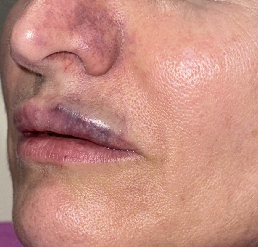
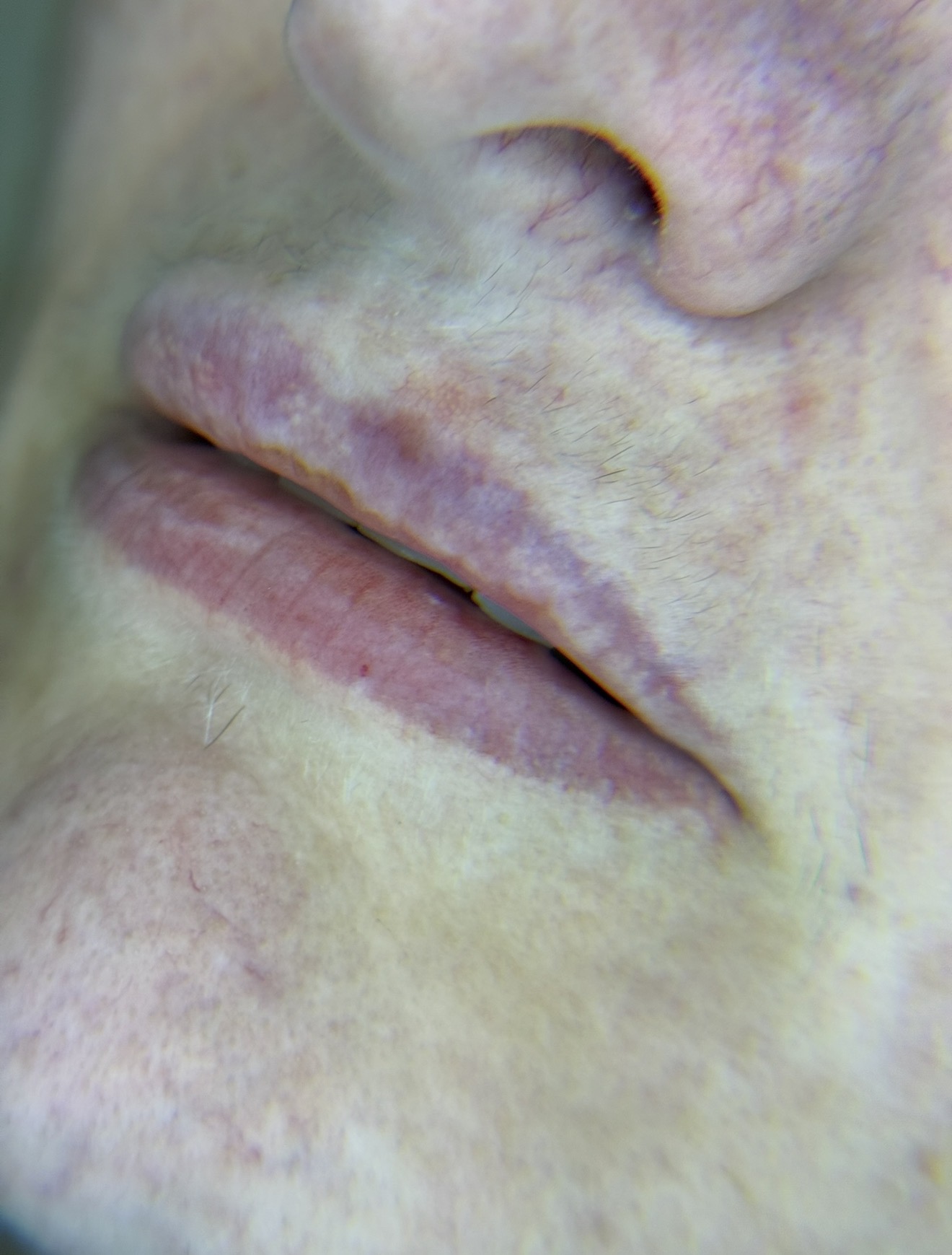
Understanding Filler Complications
Most dermal filler treatments are completed without serious complications. Mild swelling, tenderness and bruising are expected and usually settle within a few days.
However, on rare occasions, complications can occur. Some are primarily aesthetic concerns, while others require urgent medical assessment. Recognising the difference is one of the most important steps in protecting both the appearance and health of the treated area.
Filler-related complications can broadly be divided into:
- Vascular complications – where filler blocks or compresses a blood vessel, reducing blood flow to the surrounding tissue.
- Inflammatory complications – including delayed swelling, nodules and immune-mediated reactions.
- Infectious complications – which may present with increasing redness, warmth, pain or discharge.
- Product-related complications – such as filler migration, overcorrection or persistent swelling.
- Delayed complications – which may develop months or even years after the original treatment.
Each of these conditions has a different cause and requires a different management approach. Not every lump should be dissolved, not every swollen area is infected, and not every complication requires hyaluronidase.
A careful clinical assessment—and, where appropriate, high-frequency Doppler ultrasound—helps determine the most appropriate treatment rather than relying on assumptions.
EXPERIENCE • ULTRASOUND • EVIDENCE-BASED CARE
Why Patients and Doctors Refer Filler Complications to Us
01
High-Frequency Doppler Ultrasound
Rather than relying on surface appearance alone, we use high-frequency Doppler ultrasound—when clinically appropriate—to help assess filler location, surrounding tissues, and detectable blood flow. This information can guide treatment decisions and help target management more precisely.
02
Patients From Any Clinic Welcome
You do not need to have had your filler performed at our clinic.
We regularly assess patients treated elsewhere and work with the information available to determine the safest next step, without criticism of the original practitioner.
03
Evidence-Based Management
Every complication is different.
Treatment decisions are based on your history, examination findings, current clinical guidance, and—where appropriate—ultrasound assessment, rather than a routine “one-size-fits-all” approach.
04
Early Assessment Matters
Some complications become more difficult to manage as time passes.
Recognising warning signs early allows timely assessment and, when indicated, earlier intervention.
05
Ongoing Follow-up
Managing a filler complication rarely ends after the first visit.
When required, we monitor healing, repeat clinical examinations, and adjust the management plan based on your progress.
Warning Signs After Filler — Explained Simply
A little bruising, tenderness and mild swelling is normal after filler and usually settles on its own. The signs below are different — they can point to a vascular occlusion, meaning the filler has blocked or compressed a blood vessel and the tissue beyond it isn’t getting enough blood. This is the most serious complication in filler medicine, and recognising it early makes a real difference to the outcome.
Pallor (unusual paleness)
If an area of skin turns noticeably whiter than the surrounding tissue right after injection, it can mean blood flow into that area has been cut off or reduced. This is often the earliest sign of a problem and is easy to miss if it isn’t compared side-by-side with the untreated side of the face.
Tingling, numbness or disproportionate pain
Pain that feels far more intense than the injection itself would explain — or unusual tingling or numbness — can indicate that a vessel is compressed or blocked. Pain that keeps building rather than easing over the following hour is a signal to get in touch rather than wait it out.
Livedo reticularis (a mottled, net-like pattern)
This is a lace-like or net-like pattern of reddish-purple discolouration on the skin. It happens when blood is still reaching some small vessels in the area but not flowing normally through others nearby, giving the skin an uneven, blotchy appearance rather than a uniform colour. It is a recognised early sign that circulation in the area is compromised.
Grouped blisters
If small blisters appear — particularly in clusters, a few hours to a couple of days after treatment — this suggests the skin has already been short of blood supply for some time. This stage needs urgent attention rather than home care.
Skin necrosis (skin tissue starting to die)
This is the most advanced stage, where the skin turns dusky, grey, dark blue or black because the tissue has been without adequate blood supply for too long. By this point some tissue damage may already be permanent, which is exactly why the earlier signs above should never be brushed off as “just bruising.”
If you notice any of these signs — pallor, disproportionate pain or tingling, a net-like mottled pattern, grouped blisters, or skin that looks like it is starting to break down — contact us immediately rather than waiting to see if it improves.
How We Assess a Suspected Complication
Every case is assessed individually. We take a full history of what was injected, when, where and how much, and examine the area for colour, temperature, capillary refill, tenderness and any signs of skin injury.
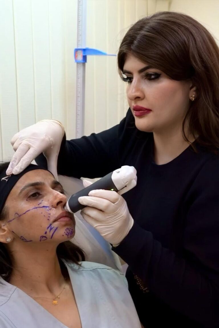
Where clinically appropriate, we use high-frequency ultrasound with Doppler to look directly at the treated area. Doppler ultrasound lets us see whether blood is actually flowing through the vessels near the injected filler, rather than relying on visual signs alone. It can help locate previously injected filler, identify fluid collections or inflammatory changes, and confirm whether a vessel is compressed — which guides exactly where and how much hyaluronidase is needed. Ultrasound adds valuable information, but it works alongside clinical judgement rather than replacing it; very small vessels are not always fully visualised.
How We Treat Vascular Complications
When a hyaluronic acid filler is responsible for a vascular occlusion, hyaluronidase is the mainstay of treatment, given promptly and reassessed repeatedly until blood flow is restored. The dose and injection pattern depend on the ultrasound findings, the product involved and how much time has passed since symptoms started — this is not a one-size-fits-all injection, and delayed or inadequate treatment is one of the main reasons outcomes can worsen.
For non-emergency concerns — unwanted filler, migration, overcorrection, or lumps and nodules — ultrasound assessment helps us confirm exactly where the product sits before deciding whether targeted dissolving, or a different approach entirely, is the right course. Not every lump should automatically be dissolved, and not every injectable responds to hyaluronidase — it will not dissolve biostimulatory fillers, permanent fillers, or your own fat.
While You’re Waiting to Be Seen
- Do not apply or inject anything not prescribed by a doctor
- Do not try to dissolve the filler yourself
- Do not press, puncture or aggressively massage the area
- Do not apply extreme heat or ice directly to the skin
- Do not delay hospital care if you have any vision, neurological, breathing or rapidly worsening symptoms
- Do take clear photographs in consistent lighting so we can track any change
What Happens Next?
━━━━━━━━━━━━━━━━━━━━━━
📞 Contact Us
━━━━━━━━━━━━━━━━━━━━━━
↓
━━━━━━━━━━━━━━━━━━━━━━
📸 Send Photos
━━━━━━━━━━━━━━━━━━━━━━
↓
━━━━━━━━━━━━━━━━━━━━━━
🩺 Medical Triage
━━━━━━━━━━━━━━━━━━━━━━
↓
━━━━━━━━━━━━━━━━━━━━━━
👩⚕️ Clinical Examination
━━━━━━━━━━━━━━━━━━━━━━
↓
━━━━━━━━━━━━━━━━━━━━━━
🔬 Ultrasound Assessment
━━━━━━━━━━━━━━━━━━━━━━
↓
━━━━━━━━━━━━━━━━━━━━━━
📋 Treatment Plan
━━━━━━━━━━━━━━━━━━━━━━
↓
━━━━━━━━━━━━━━━━━━━━━━
✅ Follow-up Review
━━━━━━━━━━━━━━━━━━━━━━
PATIENT INFORMATION & SAFETY
Frequently Asked Questions
Mild swelling and bruising are common after filler injections. However, severe pain, skin turning white, blue or purple, rapidly worsening symptoms, vision changes, or increasing skin breakdown require urgent medical assessment. If you develop vision changes, difficulty breathing, neurological symptoms or become seriously unwell, attend the nearest emergency department immediately or call 998.
Immediately if you’re worried.
Some complications benefit from early assessment, particularly if blood supply may be affected. Even if you’re unsure, it’s better to seek medical advice than wait for symptoms to worsen.
Yes.
You do not need to have had your treatment at our clinic. We regularly assess patients who have received filler elsewhere.
Please include:
• Photos
• Video
• Voice note
• Date of treatment
• Area treated
• Product if known
Yes.
Hyaluronic acid fillers may persist longer than many people expect, and delayed swelling, migration or inflammatory reactions can occasionally occur months or even years after treatment.
Yes.
Migration occurs when filler moves away from its original injection site. The cause varies depending on the product, injection technique, facial movement and tissue characteristics.
No.
Some lumps are filler.
Others are inflammation.
Others are scar tissue.
Others are infection.
Correct diagnosis comes first.
In many cases, yes.
High-frequency ultrasound can often identify the location and depth of hyaluronic acid filler and help assess surrounding tissues. However, findings depend on several factors, including the type of filler, the tissue involved and the quality of the examination.
No.
Ultrasound is an additional tool.
Medical history and physical examination remain essential.
No.
It only dissolves hyaluronic acid fillers.
It will not dissolve collagen stimulators, permanent fillers or your own fat.
Hyaluronidase temporarily breaks down naturally occurring hyaluronic acid as well as injected hyaluronic acid filler. The body continuously produces new hyaluronic acid, and treatment decisions are based on balancing the potential benefits and risks in your specific situation.
It depends.
Some patients need one treatment.
Others require several reassessments.
Ultrasound helps us understand what is happening beneath the skin before deciding on treatment. When clinically appropriate, it can assist in locating filler, assessing surrounding tissues and evaluating detectable blood flow, helping guide more targeted management.
Ultrasound can provide valuable information about blood flow in detectable vessels, but no imaging test should be relied upon in isolation. Clinical assessment remains the most important part of diagnosis.
Whenever possible, yes.
Urgent assessments are prioritised according to clinical need and appointment availability.
Usually between 30–60 minutes, depending on the complexity of the case.
Unfortunately, no.
Many complications improve with appropriate management, particularly when recognised early, but outcomes depend on the type of complication, the product involved, the anatomy affected and how much time has passed before treatment.
Most filler complications do not require surgery, although severe complications may occasionally require multidisciplinary management involving other medical specialties.
Scheduled assessment and management starts from AED 2,500. Urgent same-day complication care starts from AED 5,000. Final cost depends on the complexity of your case and is confirmed after assessment.
Medical Disclaimer
The information on this page is intended for educational purposes and should not replace an in-person medical assessment.
Every patient is different, and recommendations depend on your examination, medical history and current clinical findings.
If you experience vision changes, neurological symptoms, breathing difficulty or rapidly worsening symptoms following dermal filler treatment, attend the nearest emergency department immediately.
Still Concerned About Your Filler?
If you’re worried after a dermal filler treatment, we’re here to help.
 Call: +971 52 328 6185
 WhatsApp us with your photos, a short video, and a voice note describing your symptoms.
Patients treated at any clinic are welcome.
Filler complication management starts from AED 2,500 for a scheduled assessment and management plan. Urgent same-day complication management starts from AED 5,000, reflecting the priority triage, same-day ultrasound assessment and immediate treatment this level of care requires. The exact cost depends on the complexity of the case, the amount of hyaluronidase required, and the number of follow-up reviews needed — this is confirmed with you once you have been assessed.

What our patients think?