Home / Services / Safety & Complications, Ultrasound-Guided Treatment, Dubai Procedure Pages
Comprehensive Facial Ultrasound Mapping in Dubai
Comprehensive Facial Ultrasound Mapping in Dubai
High-frequency ultrasound of the face and neck, structured anatomical mapping, a comprehensive written report, and consultant radiologist review.
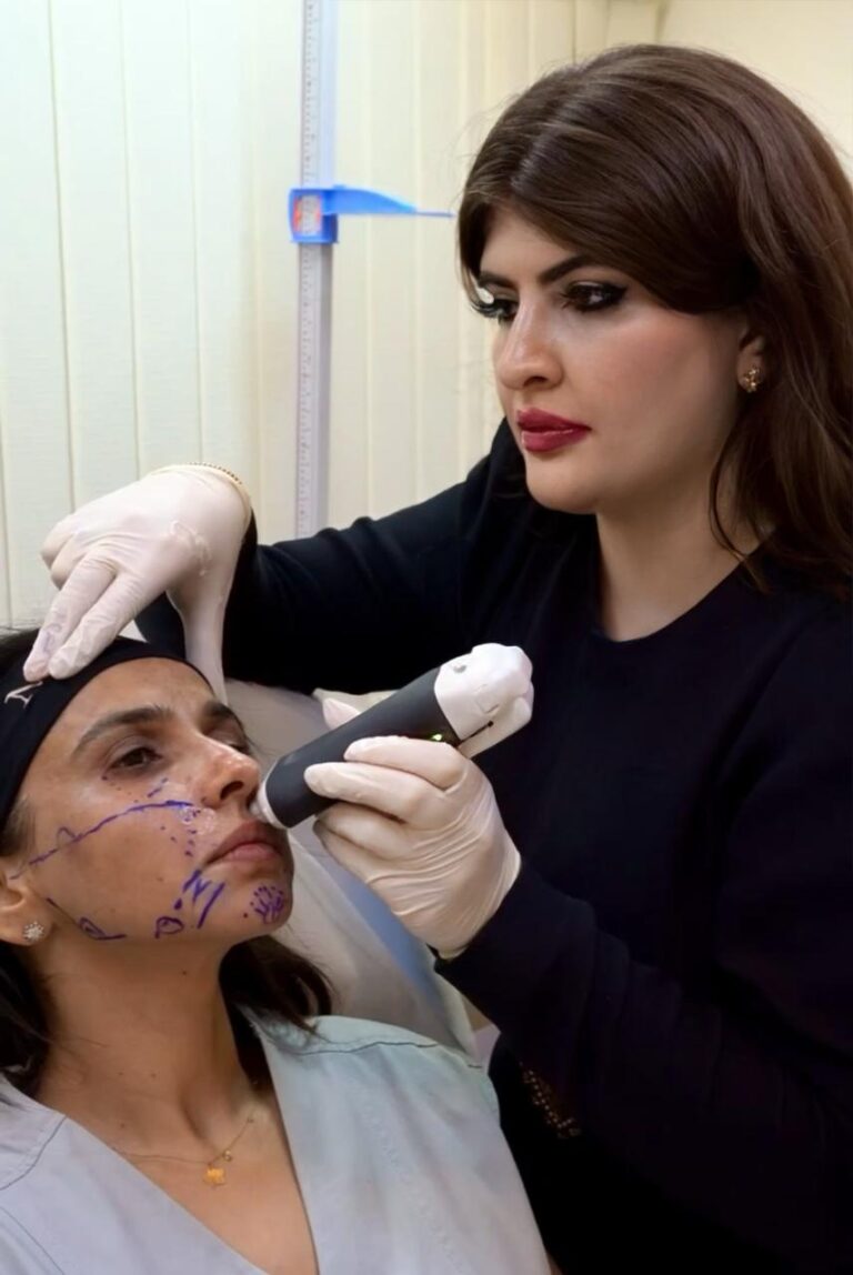
Before I treat a face that has had filler, fat grafting or threads, I want to see what is already under the skin. Not guess from the outside, and not rely on what a patient remembers being told years ago.
Comprehensive mapping examines the face and neck layer by layer: previous filler and where it sits, lumps and nodules, the quality of the skin and fat, and the vessels in the areas we plan to treat. Your findings are mapped onto anatomical illustrations and set out in a written report.
Face and neck | Colour Doppler | Illustrated report |
Systematic examination | Where clinically indicated | Maps, measurements, images |
Two ways I use ultrasound
Not every patient who needs ultrasound needs a full report. There are two different services, and you choose the one that fits.
Ultrasound as part of your treatment | Comprehensive mapping with report | |
For | A complication or concern you want me to assess and manage | Anyone who needs full documentation, complex mapping, surgical planning support or a detailed written report |
What happens | I scan the relevant area during your consultation and use ultrasound to guide treatment, for example when dissolving filler | A systematic scan of the face and neck, then mapping and an illustrated report with consultant radiologist review |
What you receive | Findings recorded in your clinical notes; your treatment plan | A comprehensive PDF report with maps, measurements, images and an impression |
cost | contact us for more info | From AED 3,150 incl. VAT |
If you have come to have a complication treated, you do not need to pay for comprehensive mapping. It is there for when the full picture, written down, is what you need.

Who comprehensive mapping is for
- You have had several treatments, in several places, and need them documented.
- You have a complex complication, or a lump or swelling that has not been explained.
- You do not know what was injected, or when.
- You have had fat grafting and something does not look or feel right.
- You are seeing a surgeon about your lower face or neck and want a detailed map to take with you.
- You want an accurate baseline before a long-term treatment plan.
What the scan shows
High-frequency ultrasound shows the soft tissues of the face in detail that no photograph or examination can. It is regarded as the first-line imaging method for identifying cosmetic fillers and their complications.1 Depending on the reason for the scan, I examine:
- Previous filler. Where it is, which layer it sits in, how much remains, and whether it has moved.
- The appearance of the material. Hyaluronic acid looks different from many permanent and semi-permanent fillers, which matters when deciding whether something can be dissolved.
- Lumps and nodules. Size, depth and, with Doppler, the blood flow around them.
- Fat grafting. Oil cysts and irregular areas after fat transfer.
- Where previous threads lie.
- Tissue quality. Thinning, thickening, fibrosis, swelling and scar tissue.
- The arteries in clinically relevant areas, mapped with colour Doppler.
- Muscles and neck structure. Such as masseter thickness on each side, and the fat above and below the platysma.
How it works
- Consultation and history. I take a detailed history of your previous treatments, symptoms, any complications and the reason for imaging.
- The ultrasound. I perform the scan myself, using Clarius high-frequency ultrasound with Doppler where clinically indicated. Each examination follows a standard protocol: still images, short video clips, measurements, Doppler findings, anatomical locations and probe orientation are recorded.
- I document the findings and prepare your face and neck maps and a preliminary imaging assessment.
- Consultant radiologist review. Your images and findings are then reviewed by Dr Abdul Rahman Alvi, who may amend, clarify or add to the interpretation.
- Your report. Once the review is complete, you receive the illustrated PDF report: anatomical maps, measurements, representative ultrasound images, findings and impression.
Dr Alvi does not need to be present at your appointment. Most examinations take approximately 30 to 45 minutes; complex cases may take longer. Your images and findings are then reviewed and the completed report is sent to you separately.
Consultant Radiologist Review
Dr Abdul Rahman Alvi, MRCS, FRCR, CCT (UK) Consultant Vascular & Interventional Radiologist Dr Alvi is a UK-trained consultant vascular and interventional radiologist. He trained at Sheffield Teaching Hospitals, completed advanced fellowships at the Royal Free Hospital, London, and holds dual accreditation from the Royal College of Surgeons of England and the Royal College of Radiologists. He has been involved in my ultrasound training, and he now reviews the imaging for comprehensive facial ultrasound mapping. His review brings a radiologist’s reading to images acquired by the doctor who will treat you. DHA licence: Consultant Interventional Radiology · 13256111-001 View Dr Alvi’s professional profile → drarjalvi.com (open in a new tab) |
(Add Dr Alvi’s photograph to the card.)
When a finding needs a procedure
Some findings need more than a report. Where a scan shows something that is best treated with an image-guided procedure, such as aspirating a cyst or fluid collection, or sclerotherapy for certain vascular lesions, Dr Alvi can carry out the procedure himself [CONFIRM: at Apogee or at his own clinic]. You are assessed first, and a procedure is only recommended when it is the right next step.
What you actually receive
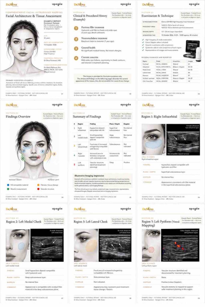
Depending on the reason for your scan, and where clinically relevant, your report may include:
- A map of previous treatments and material, with each finding numbered
- The location and tissue layer of each relevant finding
- Measurements and depth from the skin
- Compressibility and ultrasound appearance where relevant
- Doppler assessment
- Facial architecture measurements, including muscles where clinically indicated
- Tissue quality: fibrosis, thinning, thickening and swelling
- Vascular mapping of clinically relevant areas
- Submental and neck anatomy
- Representative labelled ultrasound images
- An imaging impression, with clinical or treatment considerations kept separate
- Consultant radiologist review
Not every measurement is taken in every patient. The report is built around your reason for the scan.
Example report page. Fictional patient and findings, for illustration only.
The report is written so you can understand it, with enough imaging detail to share with your surgeon, dermatologist or treating doctor.
Why it matters before treatment
Arteries in the face do not run in the same place in everyone. In a Doppler study of 80 nasolabial folds, the facial artery ran outside the fold in 31%, and its depth ranged from just under the skin to beneath the muscle.3 In 332 sides scanned near the base of the nose, the distance between the angular artery and the bone varied widely, and in 5% the artery was not present at all.4
Ultrasound also changes how filler is dissolved. When the filler can be seen, hyaluronidase can be placed into it, rather than spread across an area in the hope of reaching it.2
What ultrasound can and cannot tell you
- Ultrasound can describe the appearance and location of injected material. It cannot always identify the exact product or brand.
- Findings are written as “compatible with”, “consistent with” or “suggestive of” where that is the honest level of certainty.
- Doppler maps the vessels identified on the day of the scan. It makes treatment better informed; it does not make any injection risk-free.
- Ultrasound does not replace MRI, CT, biopsy or other tests when one of those is what the question needs.
- Sometimes the most useful result of a scan is knowing that another test or another specialist is needed.
When another investigation is needed
Depending on what the scan shows, Dr Alvi and I may recommend further imaging such as MRI or CT, an image-guided procedure, or an opinion from an appropriate surgical or medical specialist. When that happens, the report says so and explains why.
Preparing for your appointment
Come without heavy make-up if you can. No other preparation is needed.
Already had an MRI or CT? Bring it with you. If you have previous MRI, CT or ultrasound imaging relevant to the same concern, bring the images and reports with you. Relevant previous imaging may be reviewed alongside the ultrasound findings where appropriate, helping the clinical team understand how current findings relate to previous investigations. This matters most if you have previously been investigated for nodules, filler or fat-grafting complications, unknown injected material, or facial swelling. |
Also useful: previous radiology and operative reports, filler or implant records, treatment notes with product names and dates, and photographs.
Report options and price
Standard Comprehensive Report | Priority Consultant Review | |
Scan | Comprehensive face and neck, with Doppler where indicated | Same |
Radiologist review | Included | Included, prioritised |
Report delivered | Within 1 to 2 working days | 2 hours |
Price | AED 3,150 incl. 5% VAT (AED 3,000 + VAT) | AED 5,250 incl. 5% VAT (AED 5,000 + VAT) |
Priority reporting is not emergency care. Sudden pain, skin colour change, visual symptoms, rapidly spreading swelling or feeling unwell after a treatment need urgent medical attention: contact your treating doctor immediately or go to an emergency department.
Ask about availability: WhatsApp +971 52 328 6185 |
For doctors and surgeons
Plastic surgeons, dermatologists, aesthetic physicians and other clinicians are welcome to refer patients for face and neck ultrasound mapping. The report is written to be understood by the patient while keeping the imaging detail you need: findings by location and tissue layer, measurements, Doppler and labelled images.
Refer a patient / discuss a case → button: links to the clinic contact page |
Where it fits
Comprehensive mapping often comes before filler complication management, or before correcting previous work on the lips, cheeks or jawline. Neck findings feed into planning for the double chin and neck.
PATIENT INFORMATION & SAFETY
Frequently Asked Questions
No. I also use targeted ultrasound in routine assessments and ultrasound-guided treatments. Comprehensive mapping is a separate service for when you need systematic documentation, mapping of extensive previous treatment, assessment of a complex complication, baseline imaging, surgical planning support or a formal detailed report.
Not necessarily. If you want a complication assessed and managed, I scan as part of your consultation and treatment. The comprehensive report is for when you also need everything documented and reviewed by a radiologist.
Yes. Comprehensive reports are reviewed by Dr Abdul Rahman Alvi, consultant vascular and interventional radiologist.
No. I perform the scan; he reviews the images and findings afterwards.
Most examinations take about 30 to 45 minutes. The standard report follows within 1 to 2 working days.
No. Gel and a probe on the skin, no needles and no recovery time.
It can often describe whether the material looks like hyaluronic acid or something else. It cannot always identify the product or brand.
Yes, with the reports, if they relate to the same concern. They can be reviewed alongside the ultrasound findings where appropriate.
In some cases, yes. Where a finding is best managed with an image-guided procedure, such as aspiration or sclerotherapy, Dr Alvi can perform it after assessment. Otherwise we refer you to the right specialist.
Not as part of the standard examination. If a formal review of outside imaging is needed, we will discuss it with you.
Yes. It is written to be shared with your treating doctor.
It depends.
Some patients need one treatment.
Others require several reassessments.
Ultrasound helps us understand what is happening beneath the skin before deciding on treatment. When clinically appropriate, it can assist in locating filler, assessing surrounding tissues and evaluating detectable blood flow, helping guide more targeted management.
No. You can book directly, and doctors are welcome to refer.
Yes. AED 3,150 and AED 5,250 include 5% VAT.
Usually between 30–60 minutes, depending on the complexity of the case.
Unfortunately, no.
Many complications improve with appropriate management, particularly when recognised early, but outcomes depend on the type of complication, the product involved, the anatomy affected and how much time has passed before treatment.
Most filler complications do not require surgery, although severe complications may occasionally require multidisciplinary management involving other medical specialties.
Scheduled assessment and management starts from AED 2,500. Urgent same-day complication care starts from AED 5,000. Final cost depends on the complexity of your case and is confirmed after assessment.
Medical Disclaimer
The information on this page is intended for educational purposes and should not replace an in-person medical assessment.
Every patient is different, and recommendations depend on your examination, medical history and current clinical findings.
If you experience vision changes, neurological symptoms, breathing difficulty or rapidly worsening symptoms following dermal filler treatment, attend the nearest emergency department immediately.
References
- Wortsman X. Identification and complications of cosmetic fillers: sonography first. J Ultrasound Med. 2015;34(7):1163–72. doi.org/10.7863/ultra.34.7.1163
- Schelke LW, Decates TS, Velthuis PJ. Ultrasound to improve the safety of hyaluronic acid filler treatments. J Cosmet Dermatol. 2018. doi.org/10.1111/jocd.12726
- Lee W, et al. A safe Doppler ultrasound–guided method for nasolabial fold correction with hyaluronic acid filler. Aesthet Surg J. 2021;41(6):NP486–92. doi.org/10.1093/asj/sjaa153
- Franceschelli A, et al. In vivo ultrasound study of the angular artery anatomy: practical indications for the treatment of the deep pyriform space. Aesthet Surg J Open Forum. 2024;6:ojae113. doi.org/10.1093/asjof/ojae113

What our patients think?