
Home / Services / Skin Tags & Moles Treatments
Skin Tags & Moles Treatment
Removing a mole takes seconds. Deciding whether it should be removed is the part that matters.
Mole and Skin Lesion Removal in Dubai
Most people arrive having already decided. There is a mole that catches on a collar, a raised one on the cheek that photographs badly, a scatter of small skin tags on the neck that have quietly multiplied over two summers. The question in their mind is how quickly it can be done and what it costs.
Both of those have straightforward answers, and they are further down this page. But they are the second and third questions. The first is what the lesion actually is — because the laser that removes it also destroys it, and once it is gone there is nothing left to examine. That is not a reason to be nervous about mole removal. It is the reason the examination happens before the laser is switched on, and the reason some lesions are sent for a surgical opinion instead.


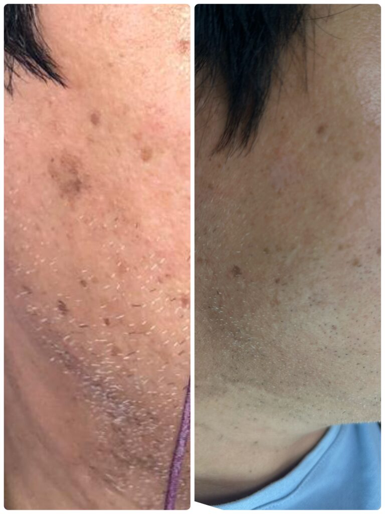
Why the lesion is identified before it is removed
An ablative laser removes tissue by vaporising it. It is precise, it can work layer by layer, and it leaves very little behind — which is exactly what makes it good at removing a mole neatly, and exactly why there is no specimen afterwards. Surgical excision is the only method that removes a lesion and preserves it for a pathologist.
So the decision to use a laser is also a decision to proceed without a laboratory report. That is entirely appropriate for a lesion that is confidently benign, and it is how most moles, skin tags and milia are dealt with everywhere in the world. It is not appropriate for a lesion that carries any question mark.
There are published cases of melanoma being found in lesions that had previously been treated with laser,2 and of melanoma arising in a lesion that had been treated as a harmless sun spot.3 There is also a well-described phenomenon in which a partly removed mole grows back with an appearance, under the microscope, that can be genuinely difficult to tell apart from melanoma.4 None of that makes laser removal a dangerous treatment. It makes the examination beforehand non-negotiable.
What the assessment involves
- Each lesion is examined with a dermatoscope, not with the naked eye. Dermoscopy is the standard tool for evaluating pigmented lesions and improves diagnostic accuracy over naked-eye examination alone.
- The history matters as much as the appearance: how long it has been there, whether it has changed, whether it itches, bleeds or crusts, and whether anyone in the family has had a melanoma.
- Where there are many moles, the lesion is judged against the others. A mole that looks unlike the rest of someone’s moles — the “ugly duckling” — earns attention even when it breaks none of the individual rules.
- If the lesion is clearly benign, it can be removed the same day. If it is not, it is not removed here.
What makes us stop Any of the following means the lesion is referred for a dermatological opinion and, usually, surgical excision with histopathology rather than laser removal: Asymmetry — one half unlike the other. An irregular, notched or blurred border. More than one colour, or colour that varies across the lesion — particularly black, blue-grey, red or white areas. A diameter that has increased, or that is larger than most of the person’s other moles. Any change: in size, shape, colour, surface or sensation. Change over weeks to months matters more than any single feature. Bleeding, crusting, ulceration, persistent itch, or a lesion that will not heal. A lesion that simply looks different from all the others on that person. A dermoscopic pattern that is not clearly that of a benign mole, whatever the lesion looks like to the eye. A mole that has changed is a reason to be examined — not a reason to have it removed faster. |
Being referred is not a delay tactic and it is not a way of avoiding a difficult case. It is the correct answer to a lesion that needs a diagnosis rather than a treatment, and it is the one thing a laser cannot provide
What can be treated here
|
Lesion |
What it is |
Usual approach |
|
Common moles (melanocytic naevi) |
Flat or raised, present for years, stable, benign on examination and dermoscopy |
Er:YAG laser ablation under local anaesthetic, one to three sessions |
|
Skin tags (acrochordons) |
Soft, skin-coloured or slightly darker tags, typically on the neck, underarms, eyelids and under the bust; often multiple |
Laser removal, usually one session; many can be done together |
|
Milia |
Small, firm, white keratin cysts, commonly around the eyes and on the cheeks |
Opened and expressed, or treated with the laser; usually one session |
|
Actinic keratoses |
Rough, scaly, sun-damage lesions — pre-malignant, not cosmetic |
Assessed first; discrete lesions can be ablated, widespread ones need field treatment or a dermatology referral (see below) |
|
Seborrhoeic keratoses |
Raised, waxy, “stuck-on” brown growths that increase with age |
Laser ablation, usually one session — confirm before publishing (§16, decision 3) |
What is not treated here
- Any lesion that is not confidently benign. These are referred for excision with histopathology.
- Lesions that have changed recently, or that the patient is worried about because they have changed.
- Large congenital moles, which are a separate problem with a separate literature and belong with a dermatologist or plastic surgeon.
- Anything ulcerated, bleeding without trauma, or failing to heal.
The laser, and why this one
Removal here is done with a Fotona erbium:YAG laser, which emits at 2,940 nm. That wavelength is absorbed by water far more strongly than the carbon dioxide wavelength is, which means the energy is spent vaporising the target rather than heating the tissue underneath it. In practice that gives a very thin zone of residual heat beneath the treated surface — and residual heat is what produces scarring. It is also what allows the lesion to be taken down in thin passes rather than in one deep one.
The public regulatory record is worth stating plainly, because very little of what is advertised about lasers in this market is checkable. The Fotona Dynamis family was cleared by the US FDA under 510(k) K101306 on 24 June 2011; the erbium:YAG dermatological indications listed in that clearance include epidermal naevi, actinic cheilitis, verrucae, skin tags, keratoses and skin resurfacing, and its general surgical indications include vaporisation, ablation and coagulation of soft tissue. The same platform carries a 1,064 nm Nd:YAG laser, cleared for photocoagulation of pigmented and vascular lesions.5
|
An honest note about indications The cleared dermatological list above names skin tags and keratoses. It does not name common melanocytic moles. Removing an ordinary benign mole with an erbium laser is a clinical judgement supported by published clinical series rather than a listed device indication — which is unremarkable in dermatology and true of a great deal of everyday practice, but it is the sort of thing worth knowing rather than discovering. What it means practically is this: the evidence for laser mole removal comes from the published outcomes, and those outcomes are set out in the next section rather than summarised as “safe and effective”. |
Laser or surgery? The trade-off, with the published numbers
A systematic review and meta-analysis published in 2026 pooled 46 studies and 4,201 treated lesions comparing surgical and laser approaches to melanocytic naevi. It is the clearest summary currently available, and the figures are worth reading before choosing a method.1
| Er:YAG laser | CO₂ laser | Surgical excision |
Complete clearance | 80.4% | 82.9% | 96.4% |
Recurrence | 14.3% | 12.9% | 2.1% |
Scarring | 2.8% | 4.8% | 21.4% |
Hyperpigmentation | 6.5% | 9.3% | 7.8% |
Overall adverse events | 11.2% | 16.4% | — |
Patient cosmetic satisfaction | 8.8 / 10 | 8.4 / 10 | — |
Specimen for the laboratory | No | No | Yes |
Read across one row and the whole decision becomes clear. Excision removes more moles definitively and almost never leaves one to come back — and leaves a visible scar in roughly one case in five. The erbium laser leaves a scar in fewer than three cases in a hundred and satisfies patients slightly more, and in exchange one lesion in seven regrows and needs a second visit. Neither is the better treatment in the abstract. They answer different questions.
The same analysis found that raised, intradermal moles recur significantly more often than flat ones (risk ratio 1.82).1 That is the single most useful predictor of how many sessions a mole will take, and it is the reason the estimate given at consultation depends on what the mole is rather than on how big it looks.
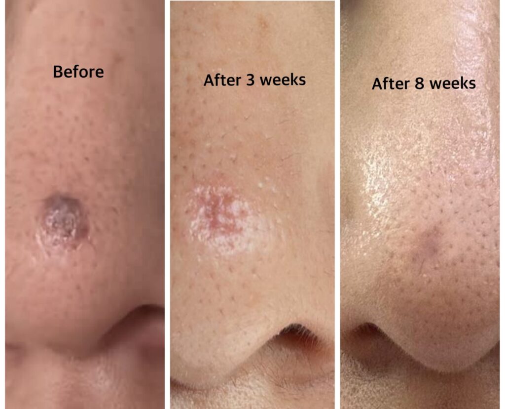
Between one and three for most moles. What decides it:
How many sessions
01
● Depth
- A flat mole sits in the upper layers and usually goes in one pass. A raised mole has a component extending deeper into the dermis, and that deeper part is what regrows if it is chased too cautiously — or what scars if it is chased too aggressively. Two conservative sessions are a better trade than one deep one.
02
● Size
- A larger surface takes longer and heals across a wider area, which raises the threshold for doing it in a single pass.
03
● Site
● Skin over the chest, shoulders and back heals less forgivingly than skin on the face and is treated more cautiously for that reason.
04
● Skin type
● Covered in the next section — and it is the factor most often left out of the estimate elsewhere.
05
even if further treatment required
A second session is not a complication and it is not a sign that the first one failed. On the published figures it is the expected course for roughly one lesion in seven, and for raised moles rather more often than that.
Skin type changes the plan — not just the risk
Dubai is a city of every Fitzpatrick type, and the same mole is not the same procedure in every one of them.
In lighter skin, healing after erbium ablation is usually quick and the treated area settles without a visible mark in most cases. In richly pigmented skin — Fitzpatrick IV, V and VI — the pigment-producing cells respond to heat and injury by producing more pigment, and the result can be a darker patch where the lesion used to be. That is post-inflammatory hyperpigmentation. It is not a scar, it is not permanent damage, and in most people it fades over weeks to months.
Two things follow from that, and the second is the one that matters:
- Pigmentation is managed, not gambled on. Strict sun protection after treatment is not optional advice — it is the single factor that most determines how quickly the area evens out. Where appropriate, a topical is added during healing.
- The treatment is deliberately more conservative. Less energy, shallower passes, more of them. That genuinely does mean darker skin types are more likely to need two or three sessions rather than one — and that is the trade being chosen on purpose, because the alternative is a faster result with a much higher chance of leaving a mark that lasts longer than the mole did.
|
Said plainly Being told a mole will take three sessions is not bad news dressed up. It means the person treating it has looked at your skin and decided not to take a risk with it. A clinic that offers every patient a single session regardless of skin type is not offering a better laser. |
Actinic keratosis — the lesion worth treating for a different reason
An actinic keratosis is not a mole and it is not a cosmetic blemish. It is a rough, dry, scaly patch — often easier to feel than to see — that develops on skin with years of accumulated sun exposure: the face, ears, scalp in thinning hair, forearms and backs of the hands. It is caused by ultraviolet damage to keratinocytes, and it sits on the pathway towards squamous cell carcinoma.
The honest arithmetic
It is common to be told that an actinic keratosis will turn into cancer. The published data are more interesting than that. In a six-year study following 7,784 actinic keratoses in 169 patients, the risk of any single lesion progressing to squamous cell carcinoma was 0.60% at one year and 2.57% at four years — low, for any one lesion. In the same study, 55% of lesions followed clinically were no longer there at one year: many regress on their own.6
And yet in that same cohort, roughly 65% of all squamous cell carcinomas arose at sites of a previously diagnosed actinic keratosis.6 Both things are true at once, and together they give the real reason to treat. It is not that this particular patch is about to become a cancer — it very probably is not. It is that actinic keratoses mark skin that has been damaged enough to produce cancers, that most such cancers emerge from that damaged field, and that there is no way to look at a dozen patches and know which one matters.
Why this is written this way The market version of this paragraph is “actinic keratosis is pre-cancer, remove it now”. It is technically defensible and it is fear-based selling, which is exactly what DHA advertising rules restrict. The version above gives the patient both numbers, which is more persuasive and happens to be true. It also sets up the next section — that treating the visible lesions is not the whole answer. |
What treatment actually involves
A discrete, well-defined actinic keratosis can be ablated with the erbium laser in the same visit as anything else being treated. Where the lesions are numerous, or the surrounding skin is diffusely sun-damaged, removing them one at a time is the wrong approach — the damage is in the field, not only in the visible patches, and field treatments exist for exactly this.
The American Academy of Dermatology’s guideline is worth quoting accurately rather than selectively: cryosurgery, topical 5-fluorouracil and topical imiquimod are all strongly recommended; photodynamic therapy is conditionally recommended; and cryosurgery is conditionally recommended over carbon dioxide laser surgery.7 Ablative laser is therefore a reasonable option for individual lesions and for resurfacing selected areas — but it is not the first-line field therapy, and recurrence after resurfacing is well documented on long-term follow-up.8
Practically, that means one of three answers at the consultation:
- A few discrete lesions on otherwise reasonable skin — ablate them, review at three to six months.
- Multiple lesions or an obviously sun-damaged field — field therapy is the correct treatment, and that is a dermatology referral or a co-managed plan, not a laser session.
- A lesion that is thickened, tender, growing, or does not behave like the others — that is a lesion for biopsy, not for the laser. Early squamous cell carcinoma can look very like an actinic keratosis.
Anyone treated for actinic keratoses needs to be seen again. This is the one thing on this page that is not a single-visit treatment, and the follow-up is the part that does the work.
Rough, scaly patches that keep coming back on the same areas?Worth having looked at properly — the assessment decides whether it is a laser problem, a field problem, or a biopsy. WhatsApp +971 52 328 6185 |
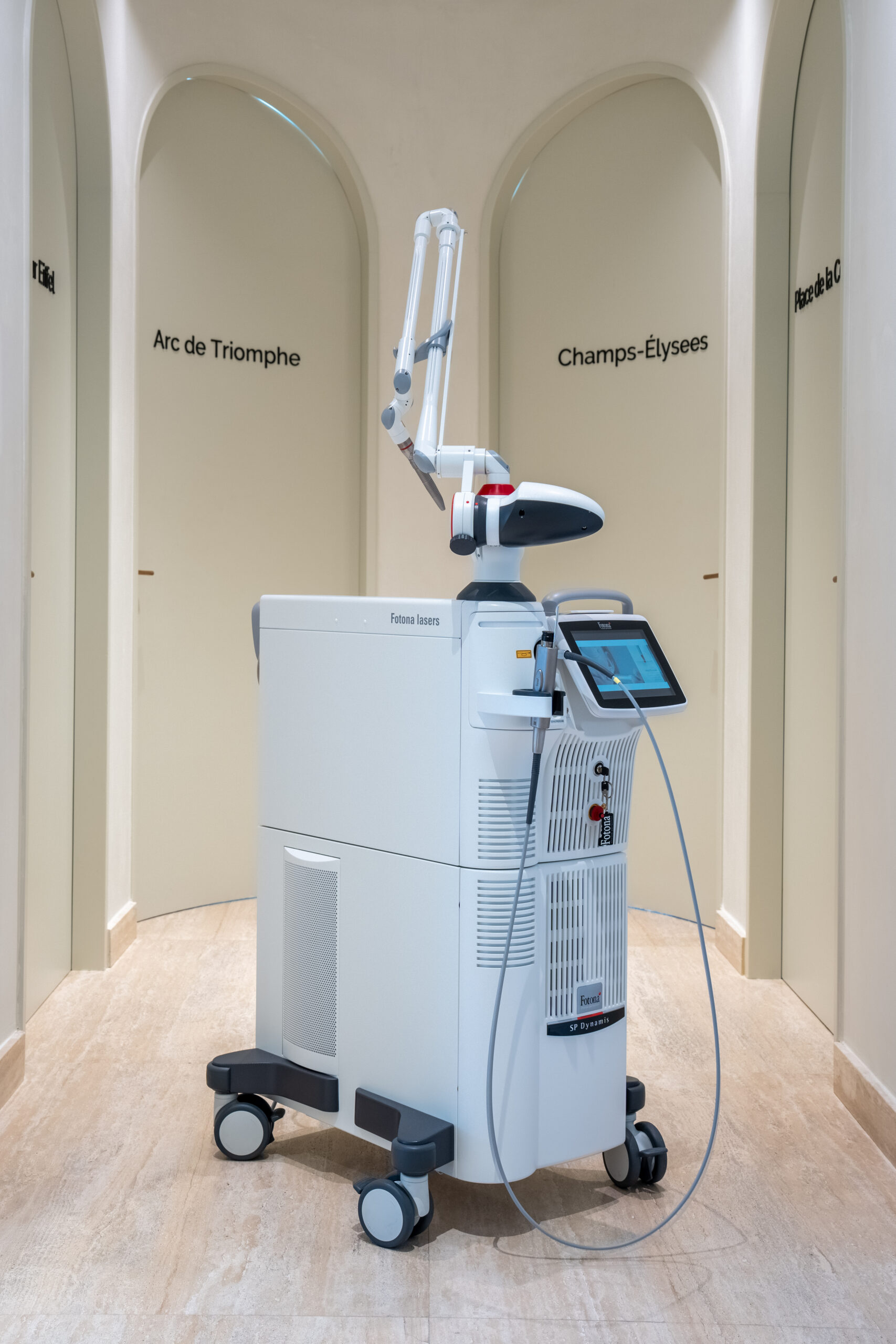
Why I Use Fotona
For the removal of suitable benign skin lesions, I use the Fotona laser because it allows precise, controlled treatment of very small areas of tissue. This is particularly useful when treating lesions such as skin tags, milia and selected benign raised lesions, where I want to target the lesion while minimising unnecessary treatment of the surrounding skin.
The treatment parameters can be adjusted according to the type, size, depth and location of the lesion, rather than using exactly the same approach for every patient. Before treatment, the lesion is assessed first; anything that appears suspicious or requires diagnostic evaluation should be referred for appropriate assessment rather than simply removed cosmetically.
Skin tags
Skin tags are soft, benign outgrowths of skin, usually on the neck, underarms, eyelids and beneath the bust — anywhere skin rubs against skin or against clothing. They are harmless. They are also one of the most satisfying things to treat, because they come away cleanly, they do not usually come back in the same spot, and a neck with thirty of them can be cleared in a single appointment.
They are removed with the same laser, under local anaesthetic where there are more than a few. Skin tags are named in the device’s cleared dermatological indications, which is worth mentioning only because so little else in laser advertising is.
One caveat, and it belongs here rather than in the small print: skin tags are common in people with insulin resistance, and a sudden increase in their number is occasionally the first visible sign of a metabolic change worth investigating. Removing them is straightforward; asking why there are suddenly so many is sometimes more useful.
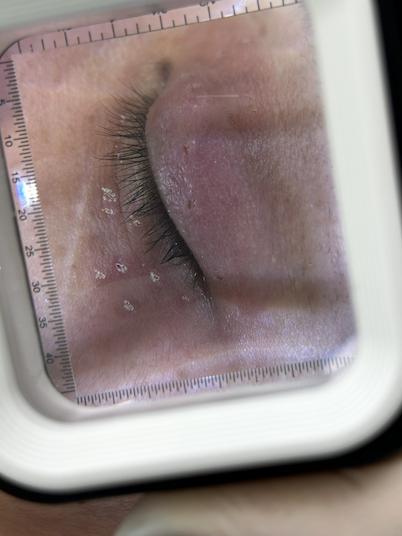
Milia
Milia are small, firm, white bumps — keratin trapped in a tiny cyst just beneath the surface, most often around the eyes and on the cheeks. They are not blocked pores, they do not respond to squeezing, and they do not resolve with a better cleanser. They need to be opened.
That is done with a fine opening and expression, or with the laser where the milium is deeper or sits somewhere delicate. It is quick and the result is immediate. Milia that keep returning in the same area are usually secondary to something — a heavy occlusive product, sun damage, or previous skin trauma — and the sensible plan is to remove what is there and change what is causing them.
What the appointment is actually like
Anaesthesia
Local anaesthetic is used for everything except the smallest milia. It is injected into the skin around the lesion, and once it has taken effect the procedure itself is not painful — the injection is the part you feel, and for most people it is comparable to a dental anaesthetic or a filler injection. For multiple lesions it is worth knowing that the anaesthetic is what takes the time; the laser part of a ten-lesion session is a matter of minutes.
During
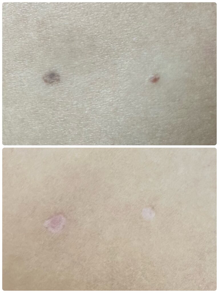
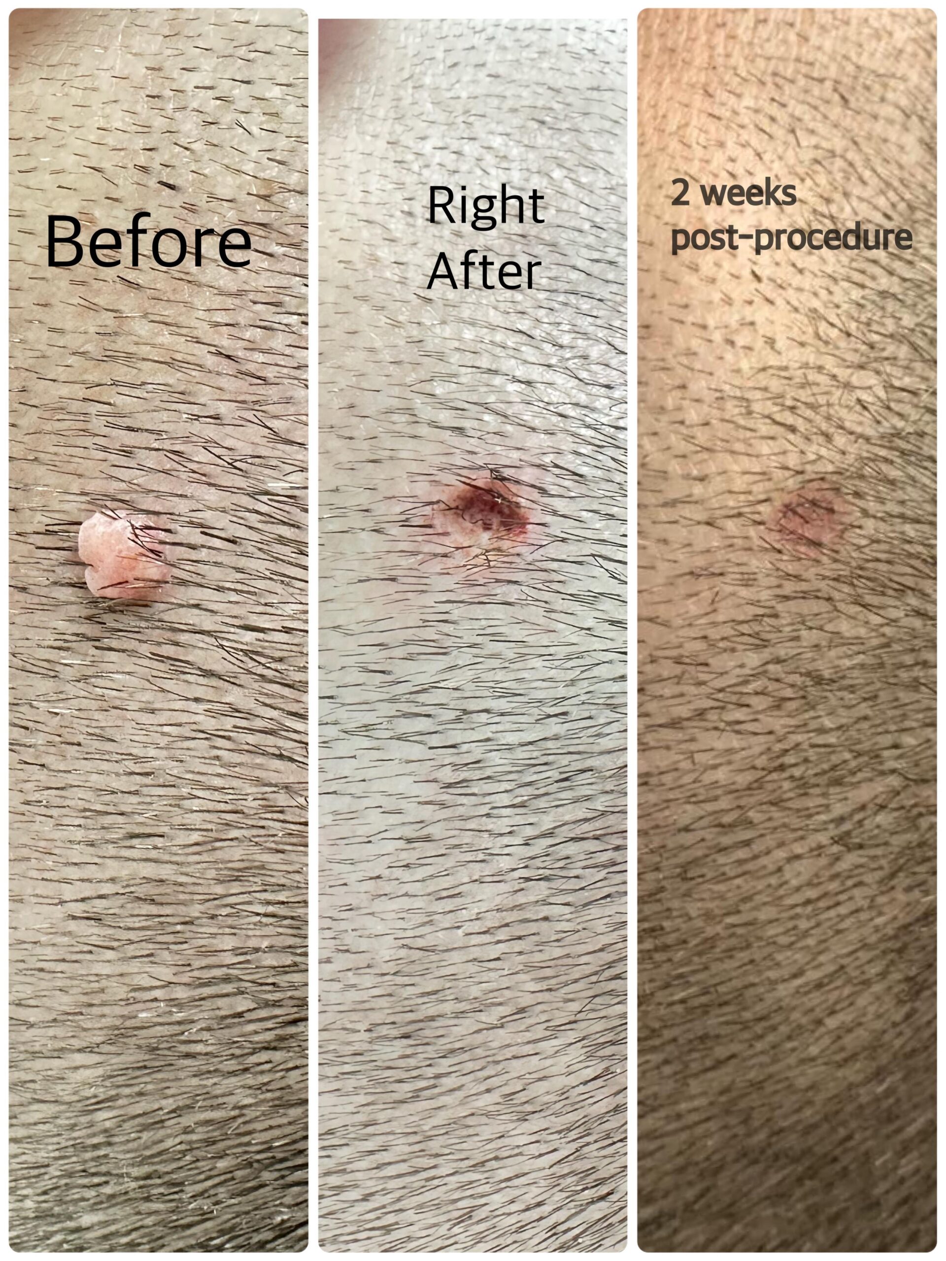
Each lesion is treated in passes, with the treated surface checked between them. There is a faint burning smell, which is normal and worth expecting. The area is left with a small shallow crater or a pinkish flat area, depending on the lesion, covered by a thin crust that forms over the following day.
Afterwards
There is remarkably little to do, and this is where a lot of clinics invent a routine. Nothing needs to be applied for the treatment to heal. The crust separates on its own, usually within seven to ten days on the face and longer on the body, and should be left alone until it does — picking it is the most reliable way to convert a clean result into a mark.
What genuinely helps:
- Sun protection. Not optional, and the single most important thing on this list — particularly in richly pigmented skin, and particularly in this climate. It is what determines whether the healed area matches the skin around it.
- For anyone who is scar-conscious, a silicone gel or silicone sheet on the healed area is the one aftercare product with a real evidence base behind it. Started after the crust has separated, not before.
- A healing cream or solution. Offered for patients who want the area to recover faster or who are treating a visible site before an event. Useful, not essential.
- No make-up over an open area until the crust has gone, and no scrubs or acids on the site while it is healing.
Normal activity, work and exercise continue as usual from the same day.
Scarring, told straight
Most people heal from erbium laser removal without a visible scar. In the pooled published data, scarring was reported in 2.8% of erbium-treated lesions, against 4.8% for CO₂ and 21.4% for surgical excision.1 Those are good odds and they are the honest reason to prefer the laser for a cosmetic removal.
They are not a guarantee, and nobody can give one. The variables that matter are the depth of the lesion, the site, how your skin has healed from injuries in the past, whether you have a tendency to keloid or hypertrophic scarring, and what you do while the crust is on. A raised mole on the chest of someone who scars readily is a different proposition from a flat mole on the cheek of someone who does not — and if that is the situation, it is better said at the consultation than discovered afterwards.
Hyperpigmentation is the more common outcome than scarring and a different thing entirely — a temporary darkening rather than a change in the skin’s structure, reported in 6.5% of erbium-treated lesions.1 It usually settles over weeks to months. Sun exposure is what makes it last longer.
And one outcome that is not a complication: the mole comes back. Roughly one in seven does, more often if it was raised. It is treated again.
Cost
Pricing is per session rather than per lesion, which is worth explaining because it changes how people plan the appointment. Most of what a session costs is the assessment, the anaesthetic and the time — not the seconds of laser energy each lesion takes. Removing ten lesions in one visit is one anaesthetic and one recovery, so it is priced as one visit.
|
Lesions treated in one session |
Price |
|
Up to 5 |
From AED 1,500 |
|
6 to 10 |
AED 2,500 |
|
More than 10 |
Discussed after consultation |
The same structure applies whether the lesions are moles, skin tags, milia or actinic keratoses, and they can be mixed within one session. If a lesion needs a second session, that is discussed at the time.
Assessment is part of the appointment. If the examination concludes that a lesion should be referred rather than removed, that is said before anything is treated.
|
Book a lesion assessment One appointment covers the examination and, where the lesions are suitable, the removal itself. WhatsApp +971 52 328 6185 |
Didn't find what you are looking for?
FAQs

The procedure itself does not, because local anaesthetic is used. What you feel is the injection of the anaesthetic — comparable to a dental injection or a filler injection, over in seconds. Once the area is numb, the removal is not painful.
Usually not. In pooled published data, scarring was reported in 2.8% of moles removed with an erbium:YAG laser, compared with 21.4% after surgical excision. Lower risk is not no risk, and depth, site, your own healing tendency and how well the crust is left alone all matter. Anyone with a history of keloid or hypertrophic scarring should say so at the consultation.
Yes, in roughly one case in seven — and more often when the mole was raised rather than flat. That is the trade-off that comes with the low scarring rate: laser removal is deliberately conservative, and being conservative means sometimes going back. A recurrence is retreated.
Because the laser vaporises it, so there is nothing left to send. This is the reason the lesion is examined carefully first, and the reason any lesion that is not confidently benign is referred for surgical excision instead — that method removes the lesion and preserves it for a pathologist.
Then it is not removed here. It is referred for a dermatological opinion and, where indicated, excision with histopathology. Features that prompt that include asymmetry, an irregular border, more than one colour, recent change in size, shape or colour, bleeding, crusting, persistent itch, or simply looking unlike the rest of your moles.
A mole that has changed is a reason to be examined, not a reason to remove it faster.
Any single actinic keratosis is unlikely to become a cancer — published follow-up puts the risk at about 0.60% at one year and 2.57% at four years, and more than half regress on their own within a year. But around 65% of squamous cell carcinomas arise at the site of a previously diagnosed actinic keratosis, and there is no way to tell which lesion will be the one. That is why they are treated, and why follow-up matters more than the removal itself.
For a few discrete lesions, it is a reasonable option. For numerous lesions or diffusely sun-damaged skin it is not first-line — guidelines strongly recommend cryosurgery, topical 5-fluorouracil and imiquimod, and conditionally recommend cryosurgery over CO₂ laser. Where the damage is in the field rather than in a few spots, the correct answer is field treatment or a dermatology referral, and that is what will be recommended.
The ones removed do not. New ones can form if whatever caused them continues — a heavy occlusive product, sun damage, or previous trauma to the area. Removing what is there and changing what is causing them is the sensible plan
Usually yes — eyelid skin tags are common and are among the most straightforward to treat. Lesions very close to the lid margin are assessed individually
The assessment is part of the appointment, so a separate visit is not usually needed. Sending a clear, unfiltered photograph beforehand is often enough to say whether the lesion is likely to be suitable. If the examination on the day concludes it should be referred rather than removed, that is said before anything is treated.