When Cheek Filler Goes Wrong:
What Ultrasound Reveals
By Dr. Shiva Faramarzi | Aesthetic & Longevity Physician, Dubai
She had been my patient for years.
She knew how I work, trusted my approach, and had always left happy. But this time, she hadn’t come to me first. She had her cheeks done elsewhere — a decision she quickly regretted.
When she came back, she was frustrated. Her cheeks looked puffier than before, heavier than they should be, and the softness she was hoping for just wasn’t there. She wanted it dissolved and redone exactly the way she remembered it.
I could see the problem in her face — but more importantly, I could see it on the ultrasound.
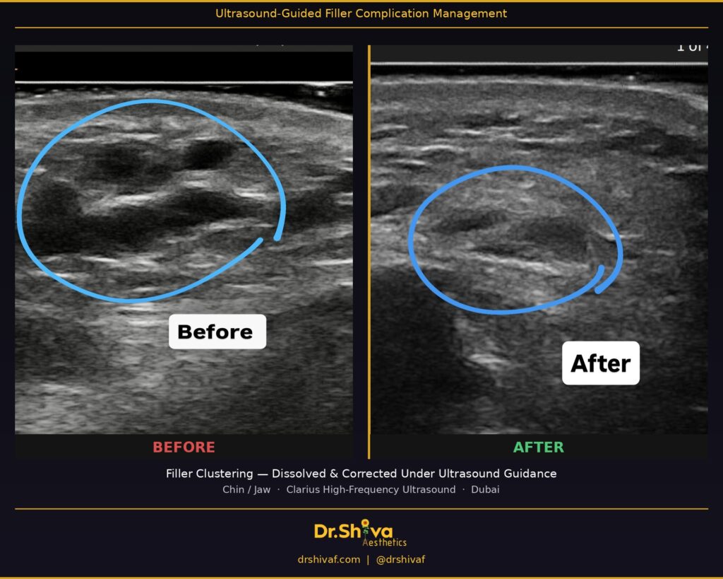
What We Found: A 2.7cm Filler Cluster
When I scanned her cheeks with the Clarius high-frequency ultrasound device, what appeared on screen told the whole story — and then some. A dense, irregular deposit of filler sitting in the wrong plane, clustered rather than distributed. The mass measured 2.7 cm.
I’ll be honest — it even formed something that looked like a smiley face on the scan. Aesthetic medicine has a sense of humour.
But behind the image is a very real lesson about what happens when injection technique is rushed:
When the plunger moves faster than the hand, filler stops sculpting and starts clustering.
Cheek filler is meant to lift and contour by distributing evenly within the correct tissue plane. When the injection speed is too fast or the placement is off, the product boluses in one place rather than integrating into the tissue. The result is exactly what she experienced — a heavy, ‘filled’ appearance rather than a lifted, natural one.
Why Ultrasound Changes Everything
Most filler dissolution in Dubai is still done ‘blind’ — the injector makes an educated guess about where the filler is sitting based on feel and surface appearance, then injects hyaluronidase in the general area.
This approach has real limitations:
- You can’t confirm how deep the filler is sitting
- You risk injecting dissolving agent into tissue that doesn’t need it
- You may dissolve unevenly, creating new asymmetries
- You can’t verify the result in real time
With ultrasound imaging, none of this is guesswork. Before I touch anything, I know:
- The exact location of every filler deposit
- The depth and plane it’s sitting in
- Whether there’s any vascular involvement nearby
- The volume and distribution — not just where filler is, but how much and in what shape
Then I dissolve under live guidance, watching hyaluronidase reach the deposit directly. And when we’re done, I scan again to confirm the tissue has cleared properly before we move to the correction.
The Correction
Once the old filler was fully dissolved and her cheeks had settled, we redid the treatment the right way — precise placement, controlled injection speed, correct plane.
The before and after ultrasound images tell the story better than any surface photo could. A clean, distributed result versus a clustered mass. That’s the difference precision makes.
What This Means for You
If your filler doesn’t look the way you hoped — or worse, looks like it’s changed over time, shifted, or feels irregular — the answer isn’t always ‘just wait.’ And it’s not always ‘dissolve and start over’ either, at least not without knowing exactly what you’re dealing with.
An ultrasound assessment gives you and your doctor a clear map before any decisions are made.
I offer ultrasound-guided filler assessment, dissolution, and correction at Apogee Clinic, JBR Dubai.
If you’re concerned about previous filler or simply want a treatment performed with full imaging precision, I’d be happy to help.